Here's what we'll cover
Here's what we'll cover
When you think of pregnancy, you probably picture a sperm and egg meeting and forming a fetus. But if something goes wrong during the process, you can end up with a mass of cells rather than a baby.
This is what’s called a molar pregnancy. It occurs due to abnormal fertilization and results in the formation of a mole rather than a fetus. The mole must be removed as it can grow and turn cancerous or permanently harm fertility.
What causes a molar pregnancy?
A molar pregnancy is a rare condition where irregular fertilization of a sperm and egg leads to the growth of cells rather than a healthy fetus. This mass of cells is called a hydatidiform mole. A hydatidiform mole can’t form into a fetus and usually results in pregnancy loss during the first or second trimester (Candelier, 2016).
To better understand abnormal fertilization, let’s see what happens during a successful one.
Most cells in our body contain two sets of DNA organized into chromosomes—one from each parent. For fertilization to happen, one sperm penetrates a DNA-containing egg. The sperm empties its DNA contents into the egg, and the two sets of genetic material fuse together forming one full set. The cell then makes copies of itself and multiplies to form a fetus (Oliver, 2020).
There are a few situations where a molar pregnancy can happen: if there are two sets of sperm (known as a partial mole), if the egg is damaged, or if the egg doesn’t have DNA to begin with (known as a complete mole) (Ning, 2019).
Partial and complete moles start out non-cancerous. If not treated properly, these masses can become invasive and evolve into a type of cancer called choriocarcinoma (Ning, 2019).
What are the risk factors for a molar pregnancy?
Like many pregnancy-related complications, scientists don’t know why molar pregnancies happen in some people and not others.
However, certain risk factors can increase the likelihood of a molar pregnancy, including (Ghassemzadeh, 2021; Murdoch, 2006):
Being of maternal age over 35 years old or younger than 20
Having previous molar pregnancies or miscarriages
Smoking cigarettes
Lack of proper nutrition (for example, old studies have found diets low in vitamin A and animal products may explain why molar pregnancies are higher in some regions of the world) (Berkowitz, 1985; Parazzini, 1988).
Having specific genes may causing multiple molar pregnancies in some individuals
Signs of a molar pregnancy
Most molar pregnancies are caught in the first trimester. Symptoms are similar to those of early pregnancy—with subtle differences.
Common signs of a molar pregnancy include:
Vaginal bleeding
Severe nausea or vomiting (often worse than typical morning sickness)
Discharge that includes grape-like cysts or clusters
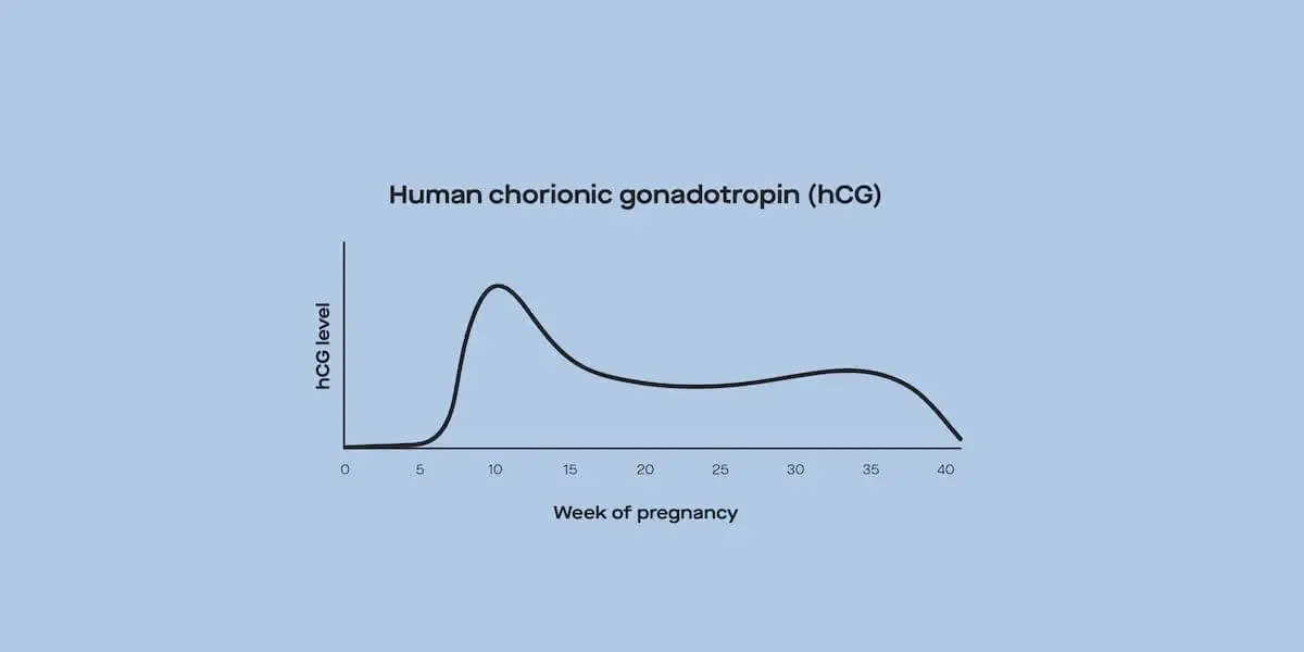
High levels of hCG: Women with complete moles have extremely high levels of hCG (human chorionic gonadotropin). This hormone that rises in early pregnancy and is used to check if you’re pregnant.
Overactive thyroid gland (hyperthyroidism)
Tremors
Rapid heartbeat
Vaginal bleeding can happen with complete and partial moles. However, bleeding caused by a complete mole is dark and more liquidy. Some may describe the blood as prune juice. No matter the appearance, it’s usually a good idea to contact a healthcare provider if you experience vaginal bleeding anytime during pregnancy. Ultimately, diagnosing a molar pregnancy requires an ultrasound (Ghassemzadeh, 2021).
How do you remove a molar pregnancy?
Once a molar pregnancy is confirmed, the standard treatment is surgically removing it to prevent complications.
The treatment is a procedure called dilation and curettage, during which medications are used to open up the cervix (the entrance to the uterus). A vacuum-like suction tool is then used to remove the molar tissue back through the cervix.
Women who don’t want to have more children can also choose to have a hysterectomy—removal of the entire uterus—to get rid of molar tissue (Cavaliere, 2009).
There have been rare cases of women with molar pregnancies giving birth. In these cases, two eggs were fertilized like with a twin pregnancy. One formed a healthy fetus, while the other formed a mole. These rare pregnancies are challenging to manage and are often complicated by severe high blood pressure (Lipi, 2019).
I had a molar pregnancy. What’s next?
After a molar pregnancy removal, it’s essential to follow up with a healthcare provider.
Depending on what kind of treatment you had, your provider will continue to monitor your hCG levels until they become undetectable. Higher hCG levels or ones that don’t go down within six months can mean there’s persistent molar tissue that needs to be treated.
It’s very uncommon, but sometimes after surgery, a small amount of molar tissue remains and continues to grow. It can then spread to other parts of the body.
Women who’ve had a molar pregnancy are at higher risk for another. But that doesn’t mean you can’t have a healthy pregnancy in the future. While you may be anxious to get pregnant, it’s best to wait until your provider gives you the go-ahead before trying again. If you become pregnant, your hCG levels start to rise again, making it difficult to know if your molar pregnancy treatment was successful (Cavaliere, 2009).
Don’t forget about or dismiss your mental health needs. You can talk to a provider during follow-up visits or seek out a therapist or counselor. Support groups also provide a community of people who have experienced similar situations as you. You can find support groups online, through social media, or your healthcare provider can give some local recommendations.
DISCLAIMER
If you have any medical questions or concerns, please talk to your healthcare provider. The articles on Health Guide are underpinned by peer-reviewed research and information drawn from medical societies and governmental agencies. However, they are not a substitute for professional medical advice, diagnosis, or treatment.
Berkowitz, R. S., Cramer, D. W., Bernstein, M. R., Cassells, S., Driscoll, S. G., & Goldstein, D. P. (1985). Risk factors for complete molar pregnancy from a case-control study. American Journal of Obstetrics and Gynecology, 152(8), 1016–1020. doi:10.1016/0002-9378(85)90550-2. Retrieved from https://sci-hub.do/10.1016/0002-9378(85)90550-2
Candelier J. J. (2016). The hydatidiform mole. Cell Adhesion & Migration, 10(1-2), 226–235. doi:10.1080/19336918.2015.1093275. Retrieved from https://pubmed.ncbi.nlm.nih.gov/26421650/
Cavaliere, A., Ermito, S., Dinatale, A., & Pedata, R. (2009). Management of molar pregnancy. Journal of Prenatal Medicine, 3(1), 15–17. Retrieved from https://pubmed.ncbi.nlm.nih.gov/22439034/
Ghassemzadeh, S., & Kang, M. (2021). Hydatidiform Mole. In StatPearls. StatPearls Publishing. Retrieved April 27, 2021 from https://pubmed.ncbi.nlm.nih.gov/29083593/
Lipi, L. B., Philp, L., & Goodman, A. K. (2019). A challenging case of twin pregnancy with complete hydatidiform mole and co-existing normal live fetus - A case report and review of the literature. Gynecologic Oncology Reports, 31, 100519. doi:10.1016/j.gore.2019.100519. Retrieved from https://pubmed.ncbi.nlm.nih.gov/31890831/
Murdoch, S., Djuric, U., Mazhar, B., Seoud, M., Khan, R., Kuick, R., … Slim, R. (2006). Mutations in NALP7 cause recurrent hydatidiform moles and reproductive wastage in humans. Nature Genetics, 38(3), 300–302. doi:10.1038/ng1740. Retrieved from https://sci-hub.do/http://dx.doi.org/10.1038/ng1740
Ning F, Hou H, Morse AN, Lash GE. Understanding and management of gestational trophoblastic disease. F1000Res. 2019 Apr 10;8:F1000 Faculty Rev-428. doi:10.12688/f1000research.14953.1. Retrieved from https://pubmed.ncbi.nlm.nih.gov/31001418/
Oliver, R., & Basit, H. (2020). Embryology, Fertilization. In StatPearls. StatPearls Publishing. Retrieved April 27, 2021, from https://pubmed.ncbi.nlm.nih.gov/31194343/
Parazzini F, La Vecchia C, Mangili G, Caminiti C, Negri E, Cecchetti G, Fasoli M. Dietary factors and risk of trophoblastic disease. American Journal of Obstetrics and Gynecology. 1988 Jan;158(1):93-9. doi: 10.1016/0002-9378(88)90785-5. Retrieved from https://sci-hub.do/10.1016/0002-9378(88)90785-5


